
Where Does Osteoarthritis Pain Come From? Part 1: The Structural Causes of Osteoarthritis Pain
- Dr Mohammad Jomaa
- Jul 5, 2026
- 8 min read
Osteoarthritis is commonly described as a disease of cartilage “wear and tear.” According to this traditional explanation, the cartilage gradually wears away, the bones begin rubbing against each other, and the joint becomes painful.
The reality is considerably more complex.
One of the most important things to understand about osteoarthritis is that the amount of pain a person experiences often does not correspond closely to the amount of structural change seen on an X-ray or MRI. Some people have advanced osteoarthritis on imaging but relatively little pain. Others experience significant pain despite having only modest changes on their scans.
This does not mean that the structure of the joint is irrelevant. It means that we need a more sophisticated understanding of which structures can actually generate pain and how they interact with the nervous system.
Osteoarthritis pain is a complex and multifaceted phenomenon. It may involve:
Structural changes within the joint
Inflammation
Changes in how the nerves process pain
Muscle weakness and altered movement
Sleep, stress and emotional health
Social and environmental influences
In medicine, we often bring these factors together using a biopsychosocial model of pain.
This article will focus specifically on the structural contributors to osteoarthritis pain. Later articles will explore the neurological, psychological and social components in greater detail.
Osteoarthritis Is a Disease of the Whole Joint
Osteoarthritis is no longer considered simply a disease of worn cartilage. It is better understood as a disorder involving the entire joint organ, including:
- Articular cartilage
- The bone beneath the cartilage
- The synovium and joint capsule
- Ligaments
- Menisci
- Tendons and muscles
- The fat pad within the knee
- The nerves and blood vessels supplying these structures
These tissues interact mechanically, chemically and neurologically. A change within one part of the joint can influence several others.
This whole-joint model helps explain why two people with apparently similar cartilage damage can have completely different pain experiences.
Cartilage Is Important — but It Cannot Directly Feel Pain
The first major message may be surprising:
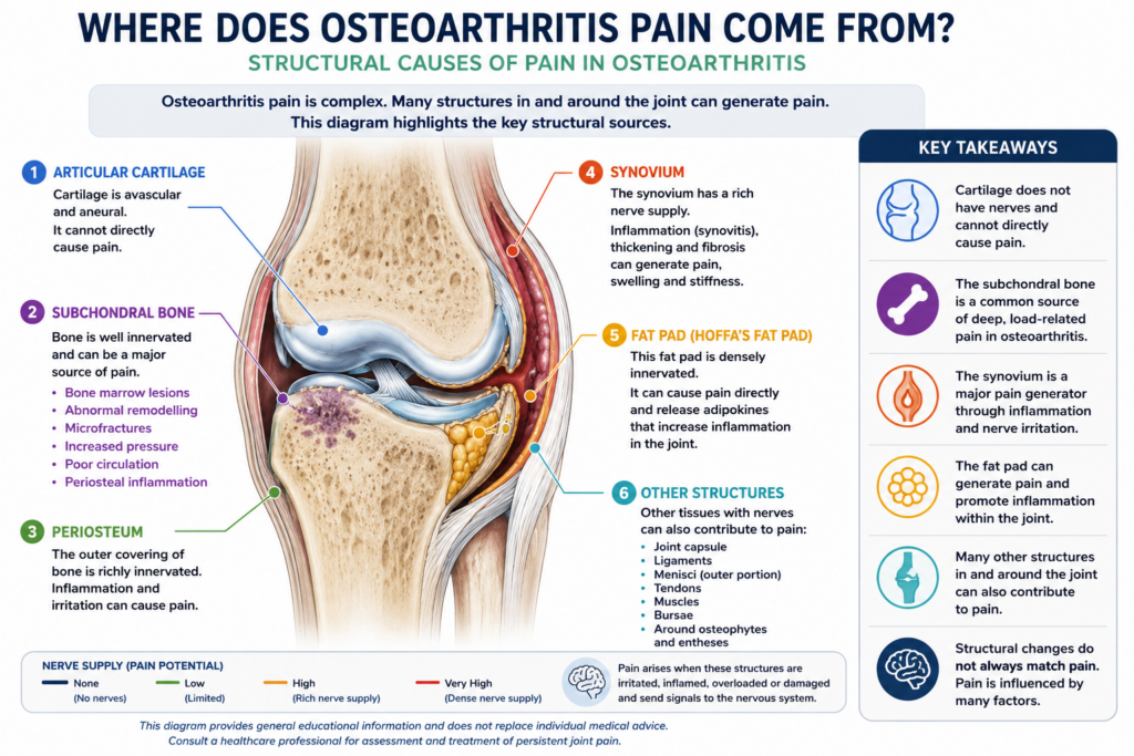
Articular cartilage does not contain nerves.
Articular cartilage is the smooth tissue covering the ends of the bones within a joint. It provides a low-friction surface, helps distribute forces and allows the joint to move smoothly.
However, articular cartilage is both:
- Avascular, meaning that it does not have its own direct blood supply
- Aneural, meaning that it does not have its own nerve supply
Because there are no pain-sensing nerves within normal articular cartilage, cartilage itself cannot directly send a pain signal to the brain.
This immediately challenges the simplistic idea that osteoarthritis pain is merely the sensation of cartilage wearing away.
That does not mean cartilage damage is unimportant. Loss of cartilage changes the way forces are distributed through the joint. It can increase loading of the underlying bone and other joint tissues. Products released during cartilage breakdown can also contribute to inflammation within the synovium.
Cartilage damage therefore contributes indirectly to pain by changing the mechanical and biological environment of the joint. But the cartilage itself is not normally the tissue producing the pain signal.
The Bone Beneath the Cartilage
Unlike cartilage, bone has both a blood supply and a nerve supply. The bone immediately beneath the cartilage is known as the subchondral bone.
This region is mechanically active. It continually adapts and remodels in response to the forces passing through the joint. In osteoarthritis, that process can become abnormal.
Possible painful changes within the subchondral bone include:
- Bone marrow lesions
- Abnormal bone remodelling
- Microfractures and trabecular injury
- Increased blood-vessel and nerve growth
- Raised pressure within the bone
- Changes in local circulation
- Irritation of the periosteum covering the outer surface of the bone
These abnormalities may stimulate the sensory nerves supplying the bone and contribute to deep, load-related joint pain.
Bone Marrow Lesions
One of the bony features most consistently associated with osteoarthritis pain is the presence of bone marrow lesions, sometimes loosely called bone marrow oedema.
On fluid-sensitive MRI sequences, these lesions often appear as bright or white areas within the bone. They are sometimes described to patients as “bone bruising” or “swelling.”
However, the term bone marrow lesion is more accurate than bone marrow oedema. These areas do not simply represent fluid. They may contain a mixture of abnormal bone remodelling, fibrosis, microdamage, altered blood vessels and other tissue changes.
In one influential study, bone marrow lesions were present in approximately 77.5% of people with painful osteoarthritic knees, compared with 30% of those whose osteoarthritic knees were not painful. Larger lesions were also more strongly associated with pain.
This is a strong association, but it is not absolute. Some people have bone marrow lesions without pain, and some people experience considerable osteoarthritis pain without detectable bone marrow lesions.
Bone marrow lesions are also not exclusively a late-stage feature. They can occur at different stages of osteoarthritis, may change in size over relatively short periods and are influenced by mechanical loading, joint alignment and the condition of the overlying cartilage.
They are therefore one part of the pain picture rather than a complete explanation.
The Periosteum and Other Bony Sources of Pain
The periosteum is the thin, highly sensitive membrane covering the outer surface of bone. Unlike cartilage, it has an extensive nerve supply.
Inflammation or mechanical irritation of the periosteum may contribute to pain, particularly around osteophytes or areas where the joint capsule and other soft tissues attach to bone.
Small areas of microdamage within the subchondral bone may also stimulate repair and remodelling. Increased pressure within the bone, altered circulation and the growth of new sensory nerves into areas of damaged subchondral bone are additional proposed pain mechanisms.
The important principle is that the painful “bone” in osteoarthritis is not simply two exposed surfaces grinding together. It is living tissue responding to altered loading, inflammation, remodelling and microscopic injury.
The Synovium: A Major Generator of Osteoarthritis Pain
The synovium is the specialised tissue lining the inside of a freely moving joint. It produces synovial fluid and helps maintain the environment required for healthy joint movement.
Unlike cartilage, the synovium has a rich nerve and blood supply.
When it becomes inflamed, a process known as synovitis, it can become an important source of pain, swelling and stiffness in osteoarthritis. MRI and ultrasound studies have repeatedly found associations between synovitis, joint effusion and greater osteoarthritis pain.
In a healthy joint, there is a balance between tissue breakdown and tissue repair. Cells within the joint produce growth factors, anti-inflammatory signals and other molecules that help maintain the joint.
In osteoarthritis, this balance may be disturbed by factors such as:
- Joint injury
- Excessive or poorly distributed loading
- Age-related cellular changes
- Metabolic factors
- Cartilage and bone breakdown products
- Repeated episodes of inflammation
The synovium responds by releasing inflammatory signalling molecules, including cytokines and other chemical mediators. These can irritate local nerve endings and make them more sensitive to movement and pressure.
Over time, the synovium may become:
- Thickened
- Inflamed
- More vascular
- Fibrotic or scarred
- Associated with increased joint fluid or effusion
These changes can contribute to aching, swelling, stiffness and pain during movement.
The synovium does not simply respond passively to damage elsewhere. It can also amplify the disease process by influencing cartilage, bone and the surrounding tissues. This is one of the reasons osteoarthritis is increasingly understood as an active biological disorder rather than an inevitable process of passive wear.
The Knee Fat Pad
Within the knee sits a normal structure known as the infrapatellar fat pad, or Hoffa’s fat pad. It lies behind the patellar tendon and is closely related to the synovium.
The fat pad helps fill space within the front of the knee and adapts its shape as the knee bends and straightens. It also has a very dense nerve supply, making it a potential source of pain when it becomes inflamed, irritated, compressed or fibrotic.
The fat pad is not simply an inert lump of stored fat. Adipose tissue is biologically active and can release signalling substances known as adipokines.
These include molecules that may influence:
- Inflammation
- Cartilage metabolism
- Synovial activity
- Blood-vessel formation
- Local nerve sensitivity
In osteoarthritis, communication between the fat pad and the neighbouring synovium may help maintain an inflammatory environment within the knee. Research has found altered expression of several adipokines in the fat pad and synovium of osteoarthritic knees.
However, it would be too simplistic to say that a larger fat pad automatically causes more pain. The biological activity and inflammatory state of adipose tissue may be more important than its size alone.
More broadly, excess body fat can influence osteoarthritis through two separate but related mechanisms:
- Mechanical loading: Greater body weight increases the forces passing through weight-bearing joints.
- Metabolic inflammation: Adipose tissue throughout the body releases biologically active molecules that may influence joint inflammation.
This helps explain why obesity is associated not only with osteoarthritis of weight-bearing joints such as the knee, but also with osteoarthritis in joints that do not carry body weight in the same way.
Other Structural Contributors
Although the bone, synovium and fat pad are important, they do not account for every structural source of osteoarthritis pain.
Other tissues containing pain-sensitive nerves include:
- The joint capsule
- Ligaments
- The outer portions of the menisci
- Tendons
- Bursae
- Muscles surrounding the joint
- The periosteum
- Areas around osteophytes
In a knee affected by osteoarthritis, for example, the capsule may become thickened or stretched by an effusion. Degenerative meniscal tissue may become displaced or irritated. Osteophytes may place tension on the capsule or surrounding soft tissues. Tendons and muscles may become overloaded as movement patterns change.
Muscle weakness can also reduce the joint’s ability to absorb and control load. This does not necessarily mean that weak muscles are themselves damaged, but it may increase the forces reaching other sensitive structures.
For this reason, pain felt “inside the joint” may sometimes arise partly from tissues immediately around it.
Why Pain and Imaging Do Not Always Match
We can now begin to understand why an X-ray does not tell us how much pain someone should have.
An X-ray primarily demonstrates bone shape, joint-space narrowing, osteophytes and other relatively established structural changes. It cannot directly show pain, nerve sensitivity, most synovial inflammation or how the joint responds during activity.
MRI provides more information, but it still offers only a structural snapshot. Abnormalities such as cartilage defects, meniscal changes and even bone marrow lesions are also commonly found in people who have no pain. MRI studies of otherwise healthy adults frequently identify structural features associated with osteoarthritis, particularly as people get older.
A scan therefore needs to be interpreted alongside:
- The patient’s symptoms
- The pattern and location of pain
- Joint swelling
- Strength and movement
- Recent changes in activity or loading
- Sleep and general health
- Neurological pain mechanisms
- The effect of pain on the person’s life
The scan is one piece of evidence. It is not the patient’s prognosis, and it should not be used in isolation to determine what the person can or cannot do.
What Does This Mean for Treatment?
Understanding where osteoarthritis pain may arise helps us move beyond the idea that treatment must simply replace lost cartilage.
Different people may have different dominant pain mechanisms.
One person may have an acutely inflamed and swollen joint with prominent synovitis. Another may have load-related subchondral bone pain. Another may have considerable muscle weakness and poor load tolerance. A different patient may have developed significant peripheral or central nerve sensitisation, meaning that the intensity of the pain can no longer be explained by the joint structure alone.
Most people will have a combination of several factors.
This is why osteoarthritis treatment usually needs to be individualised and may include:
- Education and reassurance
- Gradual modification of aggravating loads
- Strength and aerobic exercise
- Weight management where relevant
- Improving sleep and recovery
- Medication or injections in selected cases
- Addressing neurological pain sensitisation
- Managing associated psychological and social factors
- Surgery when symptoms and disability remain unacceptable despite appropriate non-operative treatment
The aim is not simply to chase every abnormality found on a scan. It is to identify the factors most likely to be driving the individual patient’s pain and loss of function.
Key Messages
Osteoarthritis pain is not simply the sensation of worn cartilage.
Articular cartilage has no nerve supply and cannot directly produce a pain signal. However, cartilage deterioration can indirectly contribute to pain by altering joint loading and stimulating inflammation.
The subchondral bone can generate pain through bone marrow lesions, abnormal remodelling, microdamage, altered circulation and increased sensory nerve activity.
The synovium is a particularly important pain-generating structure. Synovitis can contribute to pain, swelling and stiffness while also influencing the wider osteoarthritis disease process.
In the knee, the richly innervated infrapatellar fat pad may generate pain directly and may contribute to inflammation through the release of adipokines.
Other structures, including the capsule, ligaments, menisci, tendons and surrounding muscles, may also contribute.
Most importantly, structural change is only one component of osteoarthritis pain. The nervous system, physical conditioning, sleep, emotional health and wider social circumstances may all influence the final pain experience.
The next article in this series will explore the neurophysiological causes of osteoarthritis pain, including peripheral sensitisation, central sensitisation and why pain can sometimes persist or become disproportionate to the visible joint damage.
This article provides general educational information and is not a substitute for individual medical assessment. Anyone experiencing persistent, severe or unexplained joint pain should consult an appropriately qualified healthcare professional.
Selected References
- Osteoarthritis Research Society International. Understanding Your Osteoarthritis.
- Felson DT et al. The association of bone marrow lesions with pain in knee osteoarthritis. Annals of Internal Medicine, 2001.
- Neogi T. Structural correlates of pain in osteoarthritis. Clinical and Experimental Rheumatology, 2017.
- Scanzello CR and Goldring SR. The role of synovitis in osteoarthritis pathogenesis. Bone, 2012.
- Hu Y et al. Subchondral bone microenvironment in osteoarthritis and pain. Bone Research, 2021.
- Zhou S et al. Source and hub of inflammation: the infrapatellar fat pad and its interactions with articular tissues during knee osteoarthritis. Journal of Orthopaedic Research, 2022.
- Wang MG et al. The infrapatellar fat pad in inflammaging, knee joint health and osteoarthritis. npj Aging, 2024.